Microscopy is the universal diagnostic method for detection of most globally important parasitic infections.
But it's not cheap. Methods developed in well-equipped laboratories are not available at the basic levels of the health care system due to lack of resources. Researchers at the Institute for Molecular Medicine Finland, FIMM, University of Helsinki and Karolinska Institutet, Sweden, have now shown that novel techniques for high-resolution imaging and image transfer over data networks may be utilized to solve these diagnostic problems.
The team modified inexpensive imaging devices, such as a webcam selling for ten euros and a mobile phone camera, into a mini-microscope. The test sample was placed directly on the exposed surface of the image sensor chip after removal of the optics.
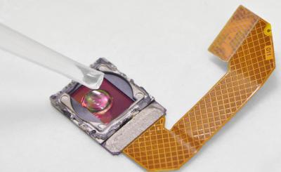
The test sample is placed directly on the exposed surface of the image sensor chip after removal of the optics.The resolution of such mini-microscopes is dependent on the pixel size of the sensor, but sufficient for identification of several pathogenic parasites. Johan Lundin Lab / Photographer: Heli Vilmi
The resolution of such mini-microscopes was dependent on the pixel size of the sensor, but sufficient for identification of several pathogenic parasites.
In their study, the researchers were able to use the mini-microscopes they constructed to yield images of parasitic worm eggs present in urine and stools of infected individuals. They first utilized this novel approach to detect urinary schistosomiasis, a severely under diagnosed infection affecting hundreds of millions, primarily in sub-Saharan Africa.
For diagnostics at the point-of-care they developed a highly specific pattern recognition algorithm that analyses the image from the mini-microscope and automatically detects the parasite eggs.
"The results can be exploited for constructing simple imaging devices for low-cost diagnostics of urogenital schistosomiasis and other neglected tropical infectious diseases", says Dr. Johan Lundin of the
University of Helsinki. "With the proliferation of mobile phones, data transfer networks and digital microscopy applications the stage is set for alternatives to conventional microscopy in endemic areas."


